TissueSurgeon
OCT-image Guided Laser Microtome
Non-Contact Precise Cutting of Tissue and Materials
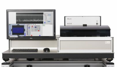
The laser microtome TissueSurgeon is a multi-purpose sectioning instrument, which enables precise and contact free cutting of biological samples and a broad range of biomaterials and materials. Based on femto-second laser technology, it can be used for sectioning, structuring or gentle extraction of tissue and materials in 2D and 3D for analysis. Fundamental limits of mechanical tissue preparation can be overcome when it comes to cutting of hard tissue, implanted tissue or difficult to cut materials.
Fields of application
- Osteology and Orthopedics (non-decalcified hard tissue and implant interface research)
- Cardiology and Cardiovascular Research and Medicine (soft tissue with biomaterials and stents, calcified plaques)
- Regenerative Medicine and Tissue Engineering (implants, scaffolds)
- Oro-facial and Dental Medicine (non-decalcified hard tissues with metal, ceramic or polymer implants)
- Oto-laryngealogy and Audiology (e.g. cochlea, implants)
- Preclinical studies from mouse to large animal models
- …
Benefits
- Nearly serial sections of non-decalcified hard tissue (e.g. bone) with minimal loss of material
- Histology of hard and soft tissue, even delicate samples (e. g. cochlea)
- Implant-tissue interface histology (e.g. dental screws, cardiovascular stents, scaffolds)
- Contact free laser cutting of tissue avoids artefacts like compressions, scratches or cracks
- Targeted and gentle isolation of site-specific samples with 3D-sections (e. g. along the implant-tissue interface of dental screws)
- Preparation of contamination and contact-free samples for (bio)-chemical analysis
- Cutting of biomaterials for tissue engineering (e.g. scaffolds, teflon, hydrogels)
- 3D-microstructuring of tissues, matrices and materials
Principle of Laser Microtomy
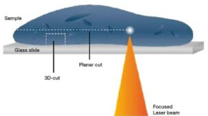
In contrast to mechanical microtomy laser microtomy is a contact free cutting method for preparation of tissue slices. Main component of TissueSurgeon is a femtosecond laser, tightly focussed into the specimen by a high-numerical aperture objective. The high intensities inside the focal volume lead to nonlinear absorption processes and finally to the disruption of the illuminated material. This effect is limited to the focal diameter of the laser pulse of approx. 1-5 µm. The whole area of the sample is scanned by the pulsed laser to perform the cutting process.
Guided Cutting by Optical Coherence Tomography
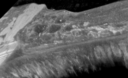
For navigation and imaging of samples or quality control of cutting, the TissueSurgeon is equipped with Optical Coherence Tomography (OCT). This provides a unique combination of two- and three-dimensional cutting and imaging, facilitating dissection and analysis of samples. Beyond, simple Multiphoton Microscopy (MPM) is an option to be integrated into the laser microtome for deeper tissue imaging.
Benefits
- Image-guided cutting allows for defined 2D-cutting and quality control
- Measurement of sample dimensions and layer thickness
- Differentiation of tissues and structures for guided cutting
- Structural information with resolution of 10 μm (OCT) up to 1μm (MPM)
- Image guided 3D sample extraction of native hard and soft tissue allows biochemical analysis of the tissue
A new dimension in laser based histology – Extra large & adjustable
| Size | Slide Size | Sample Size |
| Extra Large | 76 x 102 mm (3 inch x 4 inch) | up to 66 x 66 mm |
| Double Standard | 76 x 52 mm (3 inch x 2 inch) | up to 42 x 42 mm |
| Standard | 76 x 26 mm (3 inch x 1 inch) | up to 32 x 20 mm |
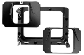
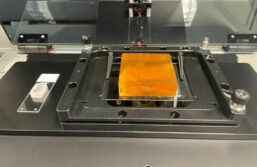
Larger slides, larger samples – More options
- Various sample holder inserts
- Sensors for automated detection of sample holder inserts
- Extra large slides available at LLS
- Customized solutions on request
Benefit from the new dimension in laser based histology, upgrade your running system.

For more information, please have a look at our product brochures or get inspired by our videos.
