Laser Based Histology
Laser Microtomy makes the difference in histology
Cost and time efficiency of preclinical studies in regenerative medicine and medical device development become more and more important to compete in the market. Histological analysis often is a mandatory, yet laborious part of the preclinical study design to get an approval. Laser microtomy can overcome fundamental limits of classical (hard tissue) microtomy and ground sectioning technology in sample preparation for histological analysis.

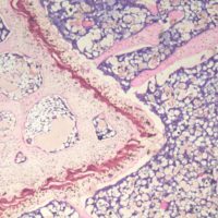
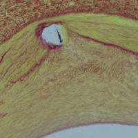
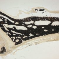
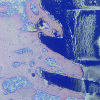
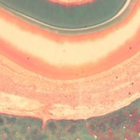
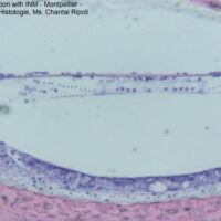
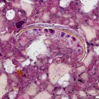
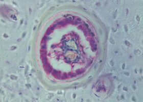
Thin sections of non-decalcified hard tissue can be generated in adequate thickness and quality as well as thin sections of implanted tissue. Even very hard non-decalcified tissue in combination with sensitive soft tissue structures can be processed without artefacts. The sections show tissue architecture and cellular details clearly. A range of histological, histochemical and immunohistochemical stains can be routinely applied.
Compared to hard tissue microtomy involving decalcification or ground sectioning technology laser microtomy reduces the processing time per sample, which increases the efficiency of lab procedures yielding in higher sample throughput.
LLS ROWIAK LaserLabSolutions GmbH is your partner for advanced histology services performed with laser microtomy.
Services
- Qualified consulting for specimen hard to cut with mechanical methods.
- Complete tissue processing from plastic embedding to cutting and staining.
- Broad range of histological, histochemical and immunohistochemical stainings.
- Development of customized protocols and staining methods for plastic embedded sections.
- Customized sectioning e.g. for synchroton or μCT analysis.
- Customized specimen preparation beyond histology, e.g. 3D cutting of native tissue
- …

Fields of Applications
- Hard Tissue
- Orthopaedics
- Oro-facial research
- Audiology
- Osteology
- Implants
- Biomaterials
- Cardio-Vascular Medicine
- Regenerative Medicine
- Tissue Engineering
- …
Embedding
For routine sectioning of non-decalcified hard and soft tissue we perform embedding in Methyl Methacrylate (MMA). For special purposes (e.g. in the case of solvent sensitive implants or biomaterials), we choose and test alternative embedding materials and protocols. Distinctive embedding and cutting protocols are developed adjusted to your requests.
Sectioning
Laser microtomy is a novel method to prepare sections for histological analysis in particular for hard tissue, implants and biomaterials.

- Fast and easy cutting of undecalcified hard tissue and a broad range of implants and biomaterials
- Semi-serial sectioning based on minimal material loss possible
- Minimization of sectioning artefacts due to contact free cutting
- Preservation of the implant-tissue interface
- Quality control of sectioning via Optical Coherence Tomography
Staining
Laser microtomy of plastic embedded samples opens new horizons in histology. A broad range of routine histological and histochemical stainings is available, e.g. Haematoxylin & Eosin, Masson Goldner Trichome, MacNeal Tetra Chrome, Tartrate-Resistant Acid Phosphatase (TRAP), …
Immunohistochemistry
Immunohistochemistry on plastic-embedded tissues including non-decalcified hard tissue opens new prospects in research of calcified tissue and tissue with implants. We offer development and application of immunohistochemistry protocols for animal (e.g. rodent, pig, rabbit, sheep, dog) tissue and human tissue covering a range of immunohistochemical markers.
Imaging by Optical Coherence Tomography
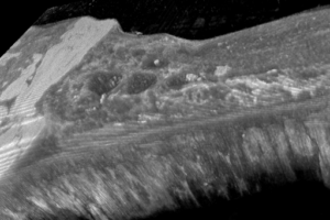
Laser microtomy with integrated OCT-imaging technology provides a unique combination of navigated two- or three-dimensional imaging and cutting, facilitating analysis and dissection of samples. It assists in the definition of cutting pattern, assessment of the cutting quality or measurement of distances. We offer
- Imaging into the depth of the sample;
- Orientation inside the sample (e.g. finding an implant,…);
- 2D-imaging and 3D-imaging of cutting pattern;
- Measurement of sample dimensions;
- Differentiation of tissues and structures for guided cutting.
Microscopy
Transmitted light microscopy and fluorescence microscopy for section quality control.
3D-Cutting for site specific tissue preparation
Beyond laser based sample preparation for histology the laser microtome enables 3D-cutting, a new way of sample collection, for e.g. biochemical analysis. The integrated optical coherence tomography allows identification of regions of interest in tissue samples and optimal positioning for cutting and isolation. This opens new dimensions of analysis in different fields such as cell biology, biomechanics or biochemical tissue analysis in regenerative medicine.
Possible Applications
- Gentle and site-specific isolation of tissue samples, e. g.
- along the implant-tissue interface, dental screws, skin implants
- for bio-mechanical tests and electrophysiological parameters (e.g. heart valves, brain, bone)
- Contamination-free trimming of sample blocks for imaging analysis i.e. for metal transition analysis by X-ray fluorescence microtomography
- …
OCT-Image of rat tibia with titanium screw implant. a) Note the bone formed after 21 days (arrow) b) a cuboid shape was cut around new formed bone, sample is ready for extraction.
Sample colletion along medical implant out of rat femur. Omar, O. Lennerås, M. Richter, H. Thomsena, P.: (2012) Laser microtome for site-specific sectioning of the interface and sub-sequent qPCR analysis (Poster Contribution)
